
 Inside Precision Medicine
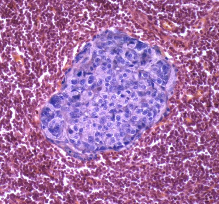
University of Cambridge researchers have developed a machine learning algorithm that facilitates accurate spatial quantification of tumor tissue on digital pathology images, potentially enabling personalized treatment decisions guided by both what the tumor looks like and what its biology reveals.
The tool, named SMMILe (Superpatch-based Measurable Multiple Instance Learning), not only matches or exceeds the performance of current state-of-the-art whole-slide image (WSI) tissue classification tools for the detection of cancer cells in tumor biopsies and surgical sections, but also predicts where the tumor lesions are located and the proportion of regions with different levels of aggressiveness.
“SMMILe stands out because it delivers precise, scene-aware quantification of tissue types acr

 Jackson Citizen Patriot
 WAND TV
 The Hacker News
 CNN Business
 KWQC
KWQC PC World
 Vogue
Vogue Fast Company Technology
 VARIETY
 Santa Maria Times Local
 The Daily Beast